Steroid Replacement in Critical Care
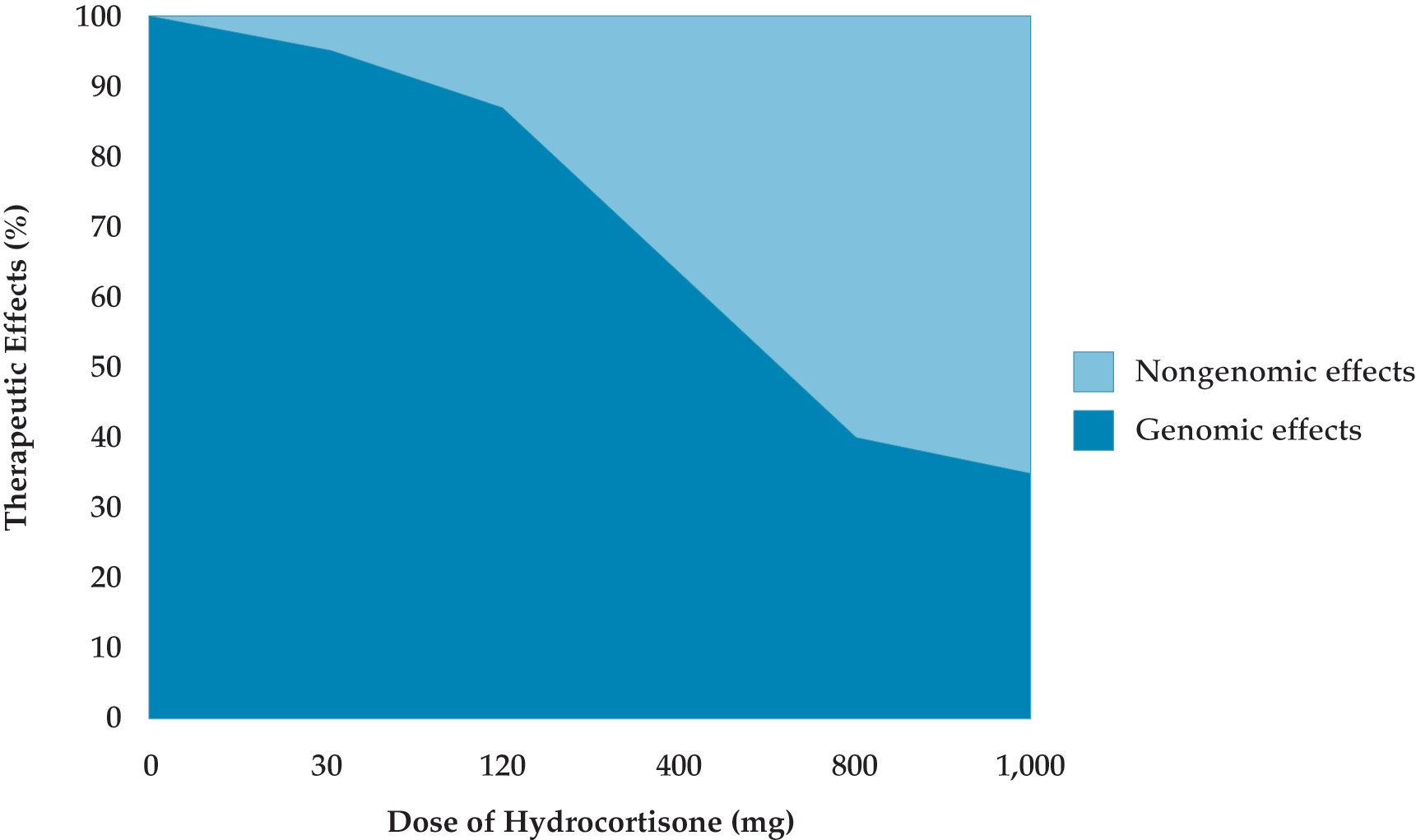
- The relative contribution of genomic and nongenomic mechanisms to corticosteroids’ therapeutic effects varies with the dose administered; the higher the dose, the greater the contribution of nongenomic effects.
- Steroid replacement may be associated with survival benefit in adults with sepsis or septic shock, acute respiratory distress syndrome, or community-acquired pneumonia.
- In sepsis, corticosteroids are best administered within the first 24 hours of management and for at least 3 days at full dose.


.png)